Lower Body Bone Diagram : Skeletal System Accessscience From Mcgraw Hill Education : Skeletal diagrams are tools used by students to learn and study all 206 bones (this number can vary from person to person) of the human body.
Lower Body Bone Diagram : Skeletal System Accessscience From Mcgraw Hill Education : Skeletal diagrams are tools used by students to learn and study all 206 bones (this number can vary from person to person) of the human body.. It consists of 5 lumbar vertebra that are numbered 1 through 5 from top to bottom i.e. Balance the weight of your head on top of your spine. It also protects several vital organs of the chest, such as the heart, aorta, vena cava, and. Your lower back (lumbar spine) is the anatomic region between your lowest rib and the upper part of the buttock. Teachme anatomy part of the teachme series the medical information on this site is provided as an information resource only, and is not to be used or relied on for any diagnostic or treatment purposes.
One of the body's largest and longest nerves is the sciatic nerve. This diagram depicts abdominal regions areas lower.human anatomy diagrams show internal organs, cells, systems, conditions, symptoms and sickness information and/or tips for healthy living. The sternum, commonly known as the breastbone, is a long, narrow flat bone that serves as the keystone of the rib cage and stabilizes the thoracic skeleton. The lower leg extends from the knee to the ankle. Balance the weight of your head on top of your spine.
Bones Of The Lower Limb Anatomy And Physiology Lower Limb Anatomy And Physiology Anatomy from i.pinimg.com Bone diagram forehead (frontal bone) nose bones (nasals) cheek bone (zygoma) upper jaw (maxilla) lower jaw (mandible) breast bone (sternum) upper arm bone (humerus) lower arm bone (ulna) thigh bone (femur) collar bone (clavicle) toe bones (phalanges) ankle bones. They hold up your body, and along with your muscles, keep you moving. The lower leg extends from the knee to the ankle. Related posts of muscles of the lower back and hip diagram muscle and bone anatomy. The long bones of the body contain many distinct regions due to the way in which they develop. Other sesamoid bones can form in the joints of the hands and feet, but are not present in all people. Skeletal diagrams are tools used by students to learn and study all 206 bones (this number can vary from person to person) of the human body. Because of the important organs situated in the abdominal area, many health concerns stem.
Key bones in the abdominal area include the base of the ribcage and the lumbar spine in the lower back.
In this image, you will find anatomy of lower extremity leg artery supplement diagram, celiac trunk, common hepatic a., superior mesenteric a., deep circumflex iliac a., external iliac a., superficial epigastric a. One of the body's largest and longest nerves is the sciatic nerve. The l5 vertebra is connected to the top of. Together, they form the part of the pelvis called the pelvic girdle. This diagram depicts abdominal regions areas lower.human anatomy diagrams show internal organs, cells, systems, conditions, symptoms and sickness information and/or tips for healthy living. Femur (2) tibia (2) fibula (2) patella (2) tarsals (14) metatarsals (10) phalanges (28) total number of bones=60. Also called the shin bone, the tibia is the longer of the two bones in the. Your body organs range from your brain, heart, liver, skin, lungs, kidneys, intestines, stomach, bladder, etc. This is the longest bone in the human body, and is also known as the thigh bone. Do you ever wonder what the major organs of the body are and. This curve, called lordosis, helps to: Related posts of muscles of the lower back and buttocks diagram muscle anatomy pics. There are two hip bones, one on the left side of the body and the other on the right.
Balance the weight of your head on top of your spine. The muscles of the lower back help stabilize, rotate, flex, and extend the spinal column, which is a bony tower of 24 vertebrae that gives the body structure and houses the spinal cord. Skeletal system diagrams are illustrations of the human skeleton, used mostly for educational purposes or in presentations. Here we will attempt to provide a brief overview of lumbar spinal anatomy. Because of the important organs situated in the abdominal area, many health concerns stem.
The Appendicular Skeleton Of Human Body Online Science Notes from onlinesciencenotes.com The patella and the pisiform bone of the carpals are the only sesamoid bones that are counted as part of the 206 bones of the body. The back supports the weight of the body, allowing for flexible movement while protecting vital organs and nerve structures. Also called the shin bone, the tibia is the longer of the two bones in the. It also protects several vital organs of the chest, such as the heart, aorta, vena cava, and. Other sesamoid bones can form in the joints of the hands and feet, but are not present in all people. The l5 vertebra is connected to the top of. Daniel nelson on june 5, 2018 8 comments ! In all, there are believed to be 80 organs in your body, all serving different functions and uses.
Related posts of muscles of the lower back and buttocks diagram muscle anatomy pics.
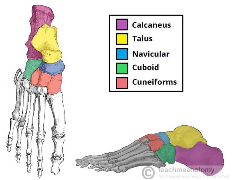
This curve, called lordosis, helps to: Your body organs range from your brain, heart, liver, skin, lungs, kidneys, intestines, stomach, bladder, etc. The tibia (shin bone) is the medial bone of the leg and is larger than the fibula, with which it is paired (figure 6.52). The sternum, commonly known as the breastbone, is a long, narrow flat bone that serves as the keystone of the rib cage and stabilizes the thoracic skeleton. The vertebral column of the lower back includes the five lumbar vertebrae, the sacrum, and the coccyx. The back supports the weight of the body, allowing for flexible movement while protecting vital organs and nerve structures. This diagram depicts bones in the lower leg 744×981.human anatomy diagrams show internal organs, cells, systems, conditions, symptoms and sickness information and/or tips for healthy living. Key bones in the abdominal area include the base of the ribcage and the lumbar spine in the lower back. The knee joint is the largest joint in the body and is primarily a hinge joint, although some sliding and rotation occur. This diagram depicts anatomy of the lower leg achilles tendon.human anatomy diagrams show internal organs, cells, systems, conditions, symptoms and sickness information and/or tips for healthy living. The bones of the leg are the femur, tibia, fibula and patella.the foot bones shown in this diagram are the talus, navicular, cuneiform, cuboid, metatarsals and calcaneus. The lower leg contains two major long bones, the tibia and the fibula, which are both very strong skeletal structures. The muscles of the lower back help stabilize, rotate, flex, and extend the spinal column, which is a bony tower of 24 vertebrae that gives the body structure and houses the spinal cord.
The anatomy of the lumbar spine is quite complex. Posteriorly the body is connected to a thin ring of bone known as the arch. Other sesamoid bones can form in the joints of the hands and feet, but are not present in all people. Evenly distribute weights from your upper body into the lower extremities. It descends from the sacral plexus through the buttocks and into the thighs to supply nerve impulses to and from the muscles and skin in the hip joints and thighs, the lower legs, feet and most of the skin below the knee.
Bones Of The Lower Limb Teachmeanatomy from teachmeanatomy.info Other sesamoid bones can form in the joints of the hands and feet, but are not present in all people. This article looks at the anatomy of the back, including bones, muscles. Evenly distribute weights from your upper body into the lower extremities. It also protects several vital organs of the chest, such as the heart, aorta, vena cava, and. The l5 vertebra is connected to the top of. The knee joint is the largest joint in the body and is primarily a hinge joint, although some sliding and rotation occur. The bones of the legs are those that make up the thigh, the lower half of the legs, and the feet. Female_anatomy_lower_body 2/3 female anatomy lower body books female anatomy lower body female anatomy lower body the internal parts of female sexual anatomy (or what's typically referred to as female) include:
The long bones of the body contain many distinct regions due to the way in which they develop.
The tibia (shin bone) is the medial bone of the leg and is larger than the fibula, with which it is paired (figure 6.52). L1, l2, l3, l4, and l5. This curve, called lordosis, helps to: 1 your spine in this region has a natural inward curve. Balance the weight of your head on top of your spine. Other sesamoid bones can form in the joints of the hands and feet, but are not present in all people. Key bones in the abdominal area include the base of the ribcage and the lumbar spine in the lower back. They hold up your body, and along with your muscles, keep you moving. Related posts of muscles of the lower back and buttocks diagram muscle anatomy pics. There are four main categories of bones: This diagram depicts bones in the lower leg 744×981.human anatomy diagrams show internal organs, cells, systems, conditions, symptoms and sickness information and/or tips for healthy living. Daniel nelson on june 5, 2018 8 comments ! The anatomy of the lumbar spine is quite complex.
Daniel nelson on june 5, 2018 8 comments ! lower body diagram. The vertebral column of the lower back includes the five lumbar vertebrae, the sacrum, and the coccyx.


0 Komentar